

Watch this video of Leslie Story.
Verdier Eye Center is excited to announce that we are now offering the cross linking procedure using Avedro KXL system, the only FDA approved cross linking device. For more information about cost and the procedure please contact our Cross Linking Coordinator, Paula Johnson, at 616-949-2001 extension 112.
Keratoconus is a medical condition which causes the cornea to become thin and weak resulting in an irregular shape or curvature. Eventually the patient’s vision becomes impaired and may interfere with day-to-day function. Cross-Linking has been proven effective in preventing disease progression and reducing corneal curvature in patients with Keratoconus.
Corneal Collagen Cross-Linking is a FDA approved surgical procedure in which the corneal epithelium (outermost layer of the cornea) is removed, Photrexa drops are applied to the surface of the cornea and the cornea is treated with ultraviolet light using a KXL cross-linking device. The goal of the treatment is to structurally stabilize the corneal tissue and delay or reduce future progression of Keratoconus. Your doctor will let you know if you are a good candidate for this procedure.
What patients need to know:
This cross-linking treatment is not recommended for women who are pregnant or lactating. It is not known whether this treatment can cause fetal harm or affect reproduction capacity. No data is present on the effects of Photrexa and Photrexa used in combination with the KXL device in relation to human milk, the effects on the breastfed infant or the effects on milk production. It is also not recommended for individuals under the age of 14 and over the age of 65 as these subjects were not included in clinical trials.
The treatment takes approximately 1 ½ hours.
The treatment is performed in an exam-room setting at Verdier Eye Center.
On the day of your treatment, have someone with you that can drive you home. You may also want to arrange to have a driver available for the 1-day post-treatment appointment as well.
There are pre-op and post-op medications used to prepare you for the treatment and help with healing after the treatment. Prescriptions of these medicines and instructions on what to do preoperatively will be given to you when the treatment is scheduled. Postoperative instructions will be given to you after the procedure. It is your responsibility to follow these instructions carefully. Medications are used to help manage pain, inflammation and help avoid infection. Medications are not always covered by insurance. It is your responsibility to find out what may or may not be covered by your insurance plan.
You will be asked to not rub the treated eye for 1 week after treatment. You may be asked to wear a protective eye shield over the eye when napping and sleeping until this cornea surface is healed.
Post-treatment symptoms:
You may experience the following symptoms related to your eye treatment… eye pain, irritation, discomfort, dryness, redness, light sensitivity, increased tearing, eye lid swelling, a temporary decrease in vision, glare/halos in vision, an increased risk of infection (while the front surface of the eye heals), possible decrease in depth perception. You may also experience non-eye related symptoms of headache, nausea and dizziness. We expect these symptoms to improve within the first few weeks after the treatment. Patients may want to arrange to have 2-3 days off of work/school. Rest and sleep are the best ways to encourage your eye to complete the initial healing stage. Use this opportunity to relax and to slow down. Sunglasses are recommended for patients experiencing light sensitivity.
A bandage contact lens may be placed in the eye at the end of your treatment. The contact lens is meant to help with pain management only (much like a bandaid over a wound) and will be removed when the doctor feels necessary. If it falls out before your exam with the doctor, do not try to re-insert it. Continue to use your post-operative medications as directed.
During the first week: You may continue normal shower/bathing routines. Try to avoid getting soap in the eye as it can cause irritation. Do not rub the eye. Avoid dusty/dirty/smoky environments for at least 1 week. Avoid swimming, hot tubs and eye makeup up to 1-month post-op.
Appointments following the treatment:
You will be instructed when you should return for follow-up exams. The normal schedule following treatment is: 1 day, 3-7 days, 1 month, 3 mos., 6 mos. and 1 year with a minimum of yearly exams thereafter. The doctors at Verdier Eye Center will instruct you on when it is safe to resume contact lens wear and when you should return to your primary eye doctor.
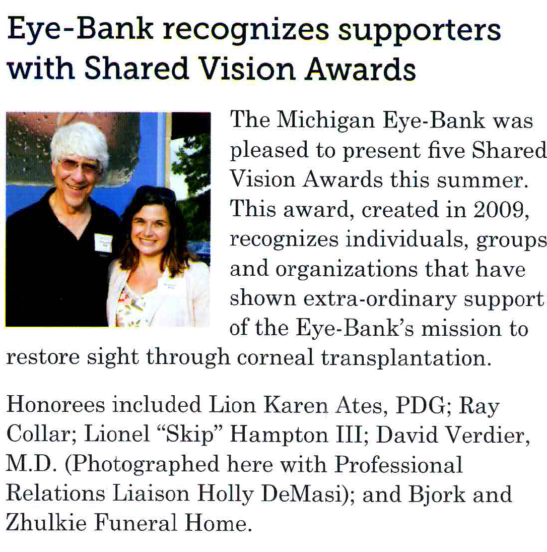
ANN ARBOR, Mich. – The Michigan Eye-Bank is pleased to announce that Grand Rapids ophthalmologist David D. Verdier, M.D., has received a Shared Vision Award.
This award, created in 2009, recognizes individuals, groups and organizations that have shown extraordinary support of the Eye-Bank’s mission to restore sight through corneal transplantation, research and education.
Dr. Verdier, who co-owns the Verdier Eye Center with Dr. Karl Siebert, is a valued partner of the Michigan Eye-Bank, and has transplanted tissue provided by the organization for more than 25 years. A resident of the Grand Rapids area since 1984, Dr. Verdier is an active member of the Eye Bank Association of America (EBAA) and a clinical professor of surgery at Michigan State University. He performed more than 125 cornea transplants in 2013.
“Dr. Verdier is considered one of the best cornea surgeons in the state of Michigan,” said Holly DeMasi, professional relations liaison at the Michigan Eye-Bank. “Over the years, we’ve received more than a few letters at the Eye-Bank from grateful recipients who are eager to share their transplant stories, thank the cornea donor families and also acknowledge what an excellent experience they have had with the whole process because of him.”
The Michigan Eye-Bank is a charitable, not-for-profit organization dedicated to the restoration of sight. It recovers, evaluates and distributes human eye tissue for transplantation. It also supports research into the causes and cures of blinding eye conditions, promotes donation awareness through public and professional education and provides humanitarian aid to people in need of corneal transplantation throughout the world. For more information, visit the Michigan Eye-Bank online at www.michiganeyebank.org or call (800) 247-7250.
Specialties:
Dr. Fox attended Ferris State University for his undergraduate and optometry studies, where he graduated with honors. He then completed a hospital based residency in ocular disease in Boston, MA, which concentrated on advanced ocular imaging methods. Prior to joining our practice, he was in private practice in coastal Maine.
Dr. Fox is a member of the American Optometric Association and the American Academy of Optometry. He has completed presentations for multiple national optometry meetings. Dr. Fox also participatedin an optometric mission trip to Honduras in 2010.
GRAND RAPIDS, Mich (January 8, 2006) – Drs. Ann Renucci and Karl Siebert recently performed two surgeries to assist a young land-mine victim from El Salvador. The patient, Mauricio Villacorta, is a 16-year-old boy who was working in a cornfield near his home in El Salvador, when a land mine exploded in his face. He was rushed to a local hospital in El Salvador where both arms were amputated. The boy suffered from facial injuries and irreparable damage to one eye. His remaining eye required extensive surgical repair, unavailable in El Salvador, to give the patient any chance of vision.
Recognizing Mauricio’s urgent medical needs, the boy was flown to the United States by Children’s Cross Connection International (CCC), an Atlanta-based non-profit medical mission organization. Through CCC, he was connected with Drs. Renucci and Siebert of Verdier Eye Center, PLC and Dr. Tom Aaberg from Associated Retinal Consultants. Dr. Renucci performed a keratoprosthesis (artificial cornea) and cornea transplant and Dr. Aaberg removed shrapnel from inside the eye. Dr. Siebert later performed a glaucoma procedure.
Following surgery, Mauricio has been able to see to recognize people and objects and ambulate independently. He is expected to regain more vision with further recuperation back home in El Salvador.